Imaging Team
We develop computational methods for image-guided surgery, medical image analysis, and 3D reconstruction. Our research advances minimally invasive spine surgery through autonomous 2D-3D fluoroscopic registration and projection-conditioned point cloud reconstruction. For Total Knee Arthroplasty (TKA), we innovate imageless navigation by integrating deep learning for precise bone morphology reconstruction and cartilage segmentation directly from intraoperative landmarks and MRI.
Our Projects
TKA (Knee)
SMART-KNEE
Imageless TKA requires realtime acquisition of patient-specific anatomical landmarks without preoperative CT/MRI. We present SMART-KNEE, a cascaded anatomy-aware reconstruction framework that generates patient-specific Femoral and Tibial bone morphology from a minimal set of intraoperatively collected anchor landmarks.
SurfMark3D
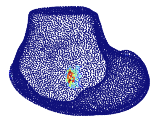
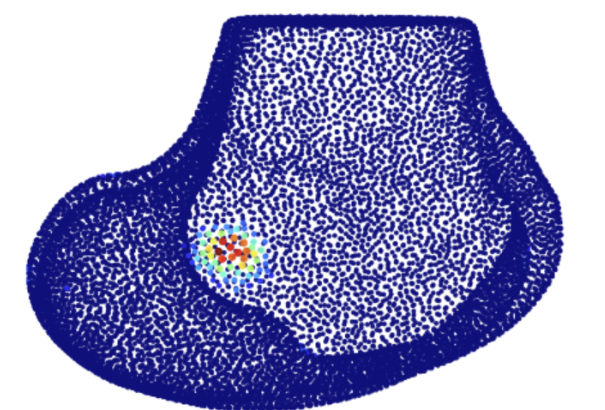
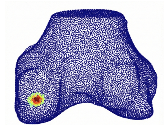
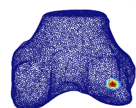
Accurate localisation of anatomical landmarks on the distal femur is a critical prerequisite for surgical planning in image-based TKA. We operate on segmented 3D surface point clouds and formulate landmark localisation as a heatmap regression task that predicts dense probability fields, achieving a Mean Euclidean Error of 1.24 mm.
MBEDNet
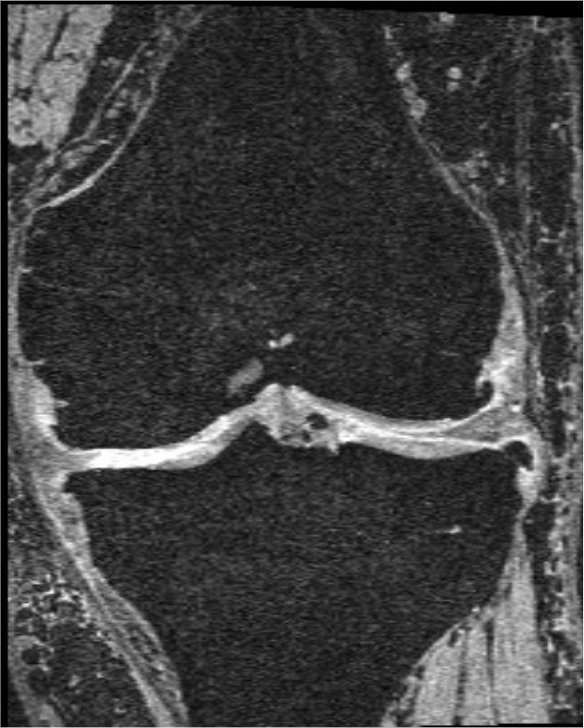
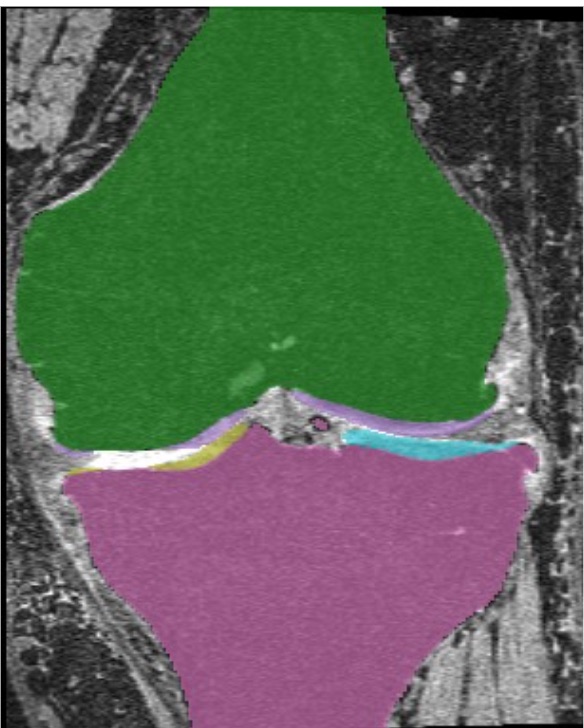
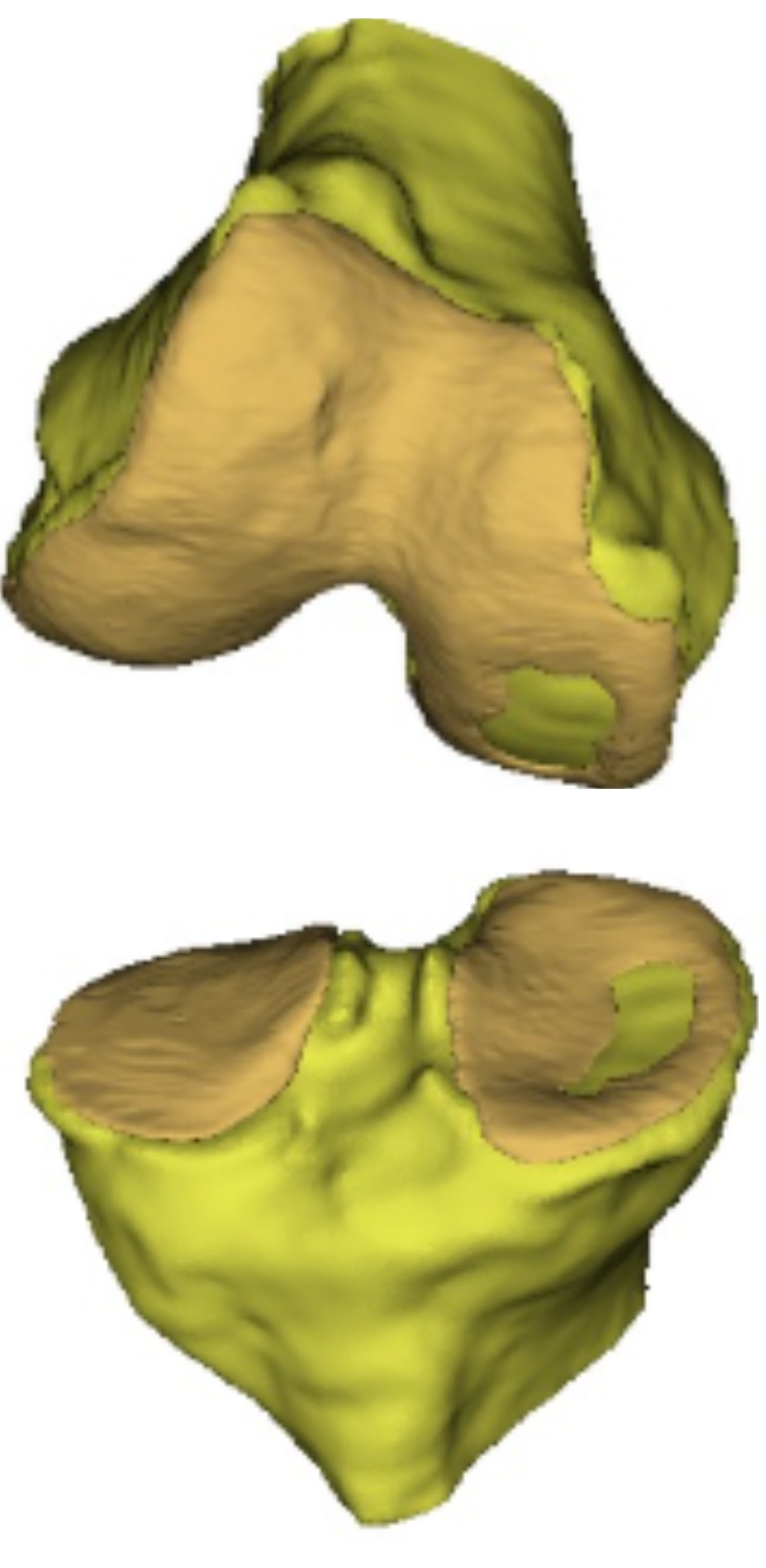
Preoperative 3D segmentation of the tibiofemoral joint guides image-based TKA. We propose Mamba Edge-Net (MBEDNet), which integrates the local feature extraction of convolution with long dependency modelling of Mamba to efficiently segment tibiofemoral anatomies, with sharp contrast between bone and cartilage.
Spine
SpineSAC
A hierarchical multi-agent Soft Actor-Critic framework that performs autonomous six-degrees-of-freedom vertebral registration in a patient-agnostic manner. Aligning CT to C-arm imagery is a foundational step in image-guided spinal surgery, and this approach reaches a target registration error of 1.45 mm on phantoms.
PPCNet
A query-refinement framework that reconstructs a dense 8,192-point cloud of spinal vertebrae from two orthogonal digitally reconstructed radiographs (DRRs) and their corresponding calibrated projection matrices. Evaluated on 105 held-out patients, it achieves a mean Chamfer distance of 1.981 mm.
Intraoperative Fluoroscopic Registration
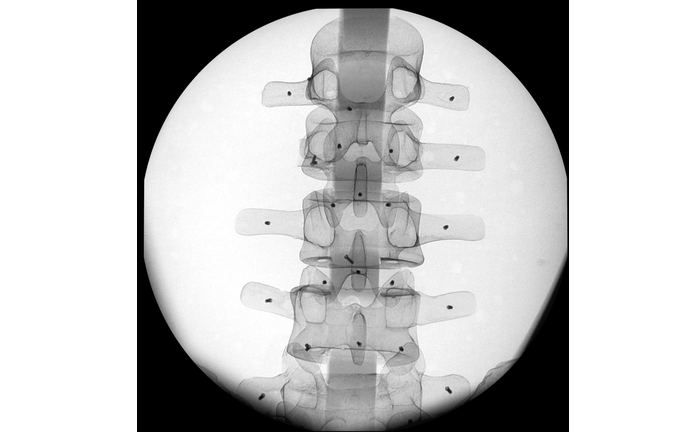
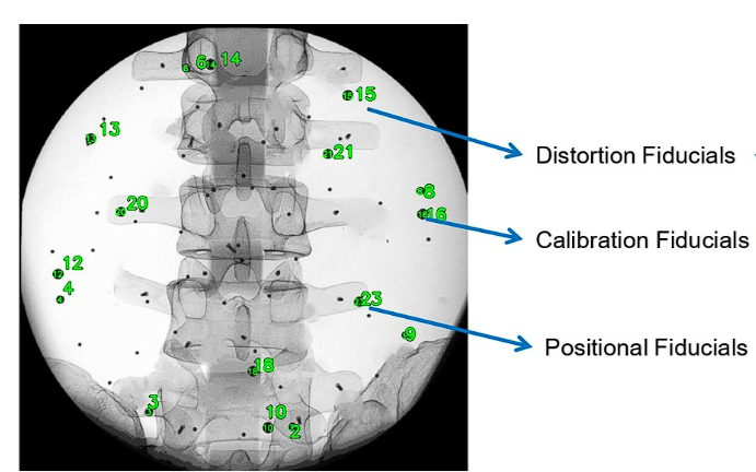
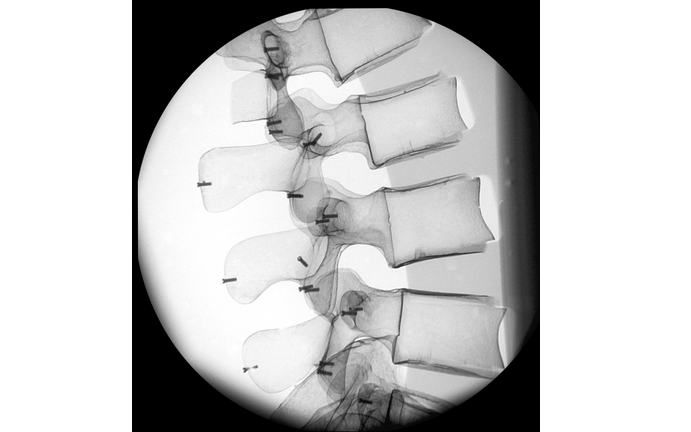
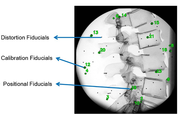
Pedicle screw placement remains a challenge in minimally invasive spine surgery. We propose a fully automatic 2D C-Arm fluoroscopic image registration system that requires only an AP and lateral image with overlaid fiducial markers from a custom-designed calibration drum, achieving sub-pixel reprojection error.